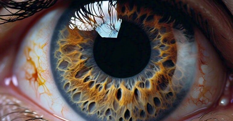
Macular Degeneration & Homoeopathy
Age-Related Macular Degeneration (AMD) is the leading cause of severe vision loss in adults over age 50. Macular Degeneration is caused by the deterioration of the central portion of the retina, the inside back layer of the eye that records the images we see and sends them via the optic nerve from the eye to the brain. The retina’s central portion, known as the macula, is responsible for focusing central vision in the eye, and it controls our ability to read, drive a car, recognize faces or colors, and see objects in fine detail.
One can compare the human eye to a camera. The macula is the central and most sensitive area of the so-called film. When it is working properly, the macula collects highly detailed images at the center of the field of vision and sends them up the optic nerve to the brain, which interprets them as sight. When the cells of the macula deteriorate, images are not received correctly.
In early stages, macular degeneration does not affect vision. Later, if the disease progresses, people experience wavy or blurred vision, and, if the condition continues to worsen, central vision may be completely lost. People with very advanced macular degeneration are considered legally blind. Even so, because the rest of the retina is still working, they retain their peripheral vision, which is not as clear as central vision.
Types of Macular Degeneration
There are two basic types of Macular Degeneration:
“dry” and “wet” .Most common “dry” macular degeneration, the tissue of the macula gradually becomes thin and stops working properly. There is no cure for dry AMD, and any loss in central vision cannot be restored.Less common, “wet” macular degeneration occurs when fluids leak from newly formed blood vessels under the macula. This leakage blurs central vision. Vision loss can be rapid and severe.
Stages
- Early AMD – Most people do not experience vision loss in the early stage of AMD, which is why regular eye exams are important, particularly if you have more than one risk factor . Early AMD is diagnosed by the presence of medium-sized drusen (yellow deposits beneath the retina).
- Intermediate AMD – At this stage, there may be some vision loss, but there still may not be noticeable symptoms. A comprehensive eye exam with specific tests will look for larger drusen and/or pigment changes in the retina.
- Late AMD – At this stage, vision loss has become noticeable.
Causes & Risk Factor
Macular degeneration is associated with aging, research suggests there also is a genetic component to the disease research has shown that oxygen-deprived cells in the retina produce a type of protein called vascular endothelial growth factor (VEGF), which triggers the growth of new blood vessels in the retina.
The normal function of VEGF is to create new blood vessels during embryonic development, after an injury or to bypass blocked blood vessels. But too much VEGF in the eye causes the development of unwanted blood vessels in the retina that easily break open and bleed, damaging the macula and surrounding retina.
Besides affecting older populations, AMD occurs females in particular. The disease also can result as a side effect of some drugs, and it seems to run in families.
Aging. The prevalence of AMD increases with age,approximately one in 14 people over the age of 40 has some degree of macular degeneration.
Obesity and inactivity. Overweight patients with macular degeneration had more than double the risk of developing advanced forms of macular degeneration compared with people of normal body weight,
Heredity. As stated above, recent studies have found that specific variants of different genes are present in most people who have macular degeneration.
High blood pressure (hypertension).
Smoking. Smoking is a major AMD risk factor
Drug side effects. Some cases of macular degeneration can be induced from side effects of toxic drugs such as chloroquine, (an anti-malarial drug) or phenothiazine anti-psychotic drugs etc.
Diagnosis
To help diagnose macular degeneration, an ophthalmologist or optometrist conduct following tests:
- Autofluorescence
- Dilated Eye Exam
- Fundoscopy or Ophthalmoscopy
- Visual Acuity Test or Eye Chart Test
- Fluorescein Angiography
- Optical Coherence Tomography (OCT)
Homeopathic Treatment
Treatments for macular degeneration depend on whether the disease is in its early-stage, dry form or in the more advanced, wet form that can lead to serious vision loss.
There is currently no known cure for Macular Degeneration, but there are things you can do to reduce your risk and possibly slow the progression once you’ve been diagnosed. For example, one can pursue lifestyle changes like dieting, exercise, avoiding smoking, and protecting your eyes from ultraviolet light.
Homoeopathy can work effectively once the pathology and symptoms has to be understood and complete case history with past medical history has to be considered then only Constitutional remedy selected with miasm can work well in ARMD.
Miasms
The exudative type of degeneration represents the sycotic miasm. The dry or atrophic type of macular degeneration more commonly represents the syphilitic miasm.
Psoric Stage – This is the earliest presentation with minimal pigmentary changes and a loss of the foveal light reflex. The foveal light reflex is produced when healthy cones reflect the light, which enters the eye. As the cells become weakened with age and disease, they loose this ability to reflect light.
Syphilitic Stage – There is more destruction of tissue with atrophy and destruction of the retina.
In last stages we can go palliative mode of treatment which can give slight relief from the symptoms.
Few Homoeopathic Remedies
- MERC SOL
Scotoma of right eye after a fall, with dimness of vision, constant flickering before eye and floating black specks. Eyes draw together. Black spots, flames, sparks before the eyes. Photophobia worse heat and glare, of fire; of foundrymen. Optic nerve and eye affection in those who work in foundries. Foggy vision.
- IODOFORMIUM
Pupils, DILATED; contract unequally, react poorly. Diplopia. Failing sight due to retrobulbar neuritis, central scotoma – partial atrophy of optic disc.
- CARBONEUM SULPHURATUM
Complete achromatopsia.Contraction of field of vision for white and blue, red and green totally absent.Central scotoma for red.Central scotoma for white and colors. Dazzled by bright light.Constant flashes and spots before the eyes. Muscae volitantes.Optic neuritis advancing towards atrophy. Arteries and veins congested. Retinal congestion ; Color-blindness.
- PLUMBUM MET
Yellow or deep bluish red sclerotics. Optic neuritis; central scotoma. Paralysis of upper lid. Sudden loss of sight after fainting. Pupils contracted. Glaucoma; from spinal affections. Profuse hot lachrymation.
NOTE – The Homeopathic medicines are selected after taking into account proper case history of the patient. The constitutional approach works best ,the Homeopathic medicines are prescribed based totality of symptoms, the and other peculiar symptoms in each individual case.
©Dr.Jitesh Sharma


Comments (4)
Edna Watson
Thanks for always keeping your WordPress themes up to date. Your level of support and dedication is second to none.
Scott James
To link your Facebook and Twitter accounts, open the Instagram app on your phone or tablet, and select the Profile tab in the bottom-right corner of the screen.